Figure 7.

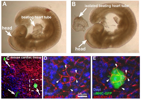
hgPSC-derived cardiac colonies can fuse with beating cardiac tissue. A-B) E9.5 fetal hearts were isolated from mouse embryos using Dumont #5 forceps. 10-15 fetal hearts were placed in one well of a 96 well plate and cMHC-GFP positive colonies were mouth pipetted into crevasses within the beating heart or simply overlaid onto the hearts. 24 hours later, hearts were analyzed live using a Leica stereoscope equipped with fluorescence. Hearts containing green areas were then fixed, stained for CNX43, and Dapi and visualized by confocal microscopy. C) Multiple GFP-positive regions were evident (arrows). D-E) Higher magnification clearly showed GFP positive cells fused to cardiac tissue via gap junctions (Arrowheads) on the same focal plane as surround heart tissue.
