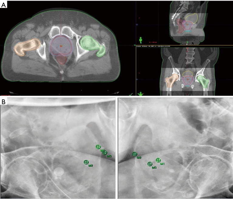
Figure 1.
IGRT consisting of CBCT (A) and stereoscopic X-ray imaging (B) with ExacTrac® at setup are used at every treatment fraction to verify the internal anatomy and intraprostatic fiducial markers. Bladder filling and the extent of rectal distension are rigorously scrutinized at the time of simulation and each treatment fraction by the attending radiation oncologist. IGRT, image-guided radiotherapy; CBCT, cone beam computed tomography.

