Figure 4.
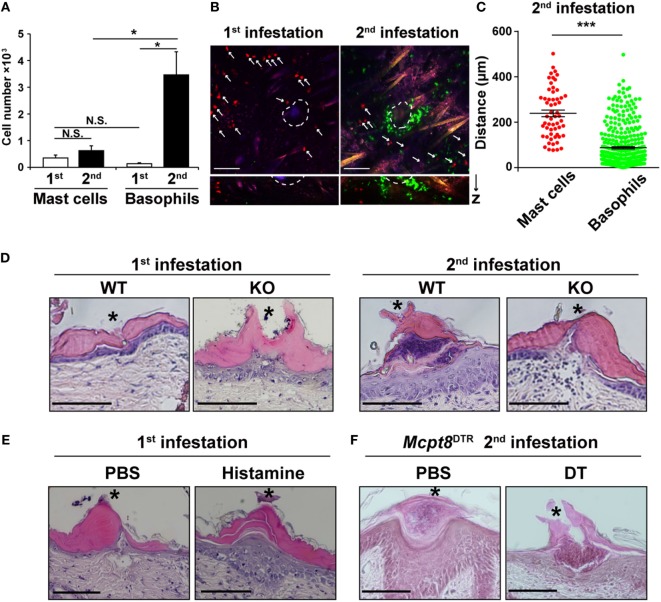
Basophils accumulate in larger numbers and at a position closer to tick mouthparts than do mast cells during the second tick infestation. (A) WT mice were infested once or twice with tick larvae. The numbers of basophils and mast cells (mean ± SEM, n = 4 each) accumulating at the second tick-feeding site on day 2 of the first or second infestation were examined by using flow cytometry. (B) Bone marrow-derived mast cell prepared from CAG-tdTomato transgenic mice were intradermally transferred into the right flank of KitW-sh/W-shMcpt8GFP mice. Recipient mice were infested once or twice with tick larvae as in Figure 3C and subjected to intravital imaging analysis of basophils (in green) and mast cells (in red, marked with white arrows) at tick-feeding sites on day 2 of the first or the second infestation. Upper pictures show the compilation of 12 horizontal images acquired from 0 to 100 μm deep under the skin. Lower panels show vertical images of upper pictures. Dashed circular lines indicate sites where tick mouthparts were inserted into the skin. Scale bars indicate 100 μm. (C) The distance of each basophil and mast cell away from the nearest tick mouthpart was measured in the images of the second tick-feeding site (mean ± SEM, 58 mast cells and 371 basophils were analyzed). (D) Skin sections of tick feeding sites in WT and histidine decarboxylase KO mice on day 2 of the first or second infestation were stained with hematoxylin and eosin. Asterisks indicate sites where tick mouthparts were inserted into the skin. Scale bars indicate 200 µm. (E) Histamine or control PBS was injected intradermally beneath the first tick-infested site once a day for 3 days, starting 1 day before the infestation. On day 2 of the first infestation, skin sections of tick feeding sites were stained with hematoxylin and eosin as in (D). (F) Mcpt8DTR mice were treated with diphtheria toxin or control PBS 1 day before the second infestation. Skin sections of tick feeding sites were stained with hematoxylin and eosin as in (D). All the data shown in (A–F) are representative of at least three independent experiments. N.S., not significant; *p < 0.05; ***p < 0.001.

