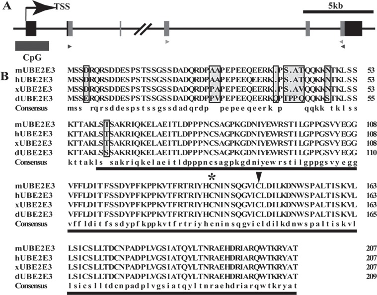
Figure 1.
UbcM2 was highly conserved in vertebrates. (A) Schematic representation of the UbcM2 gene structure (Mus musculus, chromosome 2 2D). Gray boxes represent the coding region of UbcM2. Dark rectangle demonstrates a predicted CpG island harboring a stretch of 140 CpG sites (http://genome.ucsc.edu). The black and gray triangles label the primers for specific sense and antisense probe preparation and qRT-PCR, respectively. (B) Amino acid sequence alignment of the primary sequences of mouse UbcM2 (mUBE2E3) and its orthologues in human (hUBE2E3), Xenopus laevis (xUBE2E3), D. rerio (dUBE2E3). The highly conserved UBC core domain is underlined. The conserved catalytic cysteine (145) is indicated by an arrowhead. The noncatalytic cysteine (136), which is identified as a putative redox sensor, is indicated by an asterisk.

