
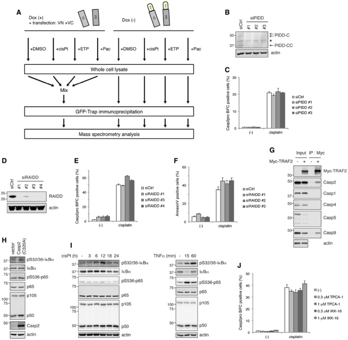
Figure EV2. Workflow of mass spectrometry analysis. Cisplatin induces caspase‐2 dimerization independently of either the PIDDosome or the NF‐κB pathway.
-
AWorkflow for mass spectrometry analysis. Casp2pro BiFC cells were treated with mock (DMSO), 20 μM cisplatin, 50 μM etoposide, or 100 nM paclitaxel in the presence of 10 μM Q‐VD(OMe)‐OPh for 24 h followed by GFP‐Trap immunoprecipitation. Note the control: Casp2pro BiFC expression was prevented with 1 μg/ml doxycycline, and unconjugated BiFC constructs (VN and VC) were co‐transfected into cells before treatment, similar to experimental samples. Control lysates were then pooled before GFP‐Trap IP. GFP‐Trap beads were subjected to on‐bead protein digestion and analyzed by mass spectrometry.
-
B, CCasp2pro BiFC cells were transfected with siRNA targeting PIDD for 48 h, and knockdown efficiency was confirmed by IB (B). PIDD siRNA transfected cells were treated with 20 μM cisplatin in the presence of 10 μM Q‐VD(OMe)‐OPh for 24 h. Caspase‐2 BiFC was assessed by flow cytometry. n = 3 independent experiments (means + s.e.m.) (C).
-
D, ECasp2pro BiFC cells were transfected with siRNA targeting RAIDD for 48 h, and knockdown efficiency was confirmed by IB (D). RAIDD siRNA transfected cells were treated with 20 μM cisplatin in the presence of 10 μM Q‐VD(OMe)‐OPh for 24 h. Caspase‐2 BiFC was assessed by flow cytometry. n = 3 independent experiments (means + s.e.m.) (E).
-
FHeLa cells were transfected with RAIDD siRNA for 48 h and then treated with 20 μM cisplatin for 24 h. Apoptosis was assessed by annexin V staining and flow cytometry. n = 3 independent experiments (means + s.e.m.).
-
GHeLa cells were transfected with Myc‐TRAF2 construct and cultured 48 h for expression. Cell lysates were prepared, followed by Myc‐Trap immunoprecipitation and IB for indicated caspases.
-
HHeLa cells were transfected with caspase‐2(C320A) construct and cultured 48 h for expression. Cell lysates were prepared, followed by IB.
-
IHeLa cells were treated with 20 μM cisplatin in the presence of 10 μM Q‐VD(OMe)‐OPh (left) or treated with 20 ng/ml TNFα (right) for indicated periods. Cell lysates were prepared and analyzed by IB.
-
JCasp2pro BiFC cells were treated with 20 μM cisplatin in the presence of 10 μM Q‐VD(OMe)‐OPh with or without indicated NF‐κB pathway inhibitors, TPCA‐1 or IKK‐16, at indicated concentrations for 24 h. Caspase‐2 BiFC was assessed by flow cytometry. n = 3 independent experiments (means + s.e.m.).
Source data are available online for this figure.