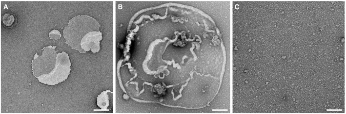
TEM image of liposomes made from E. coli polar lipid extract such as used in this work. During adsorption onto the TEM grid, liposomes fused with each other forming lipid membranes. Liposomes made from other lipids used in this work appeared very similar (not shown here).
TEM image of liposomes made from E. coli polar lipid extract and incubated with 5 μM GSDMD overnight at 37°C in the absence of caspase. None of the lipid membranes imaged (n > 50) showed the characteristic arc‐, slit‐, or ring‐like structures observed for GSDMDNterm.
TEM image of GSDMD (5 μM) incubated with caspase‐1 (0.1 μM) overnight at 37°C. No lipid or liposomes has been added to the sample.
Data information: Samples were negatively stained with uranyl acetate and imaged at 120 kV (
Materials and Methods). Scale bars, 500 nm (A) and 100 nm (B, C).