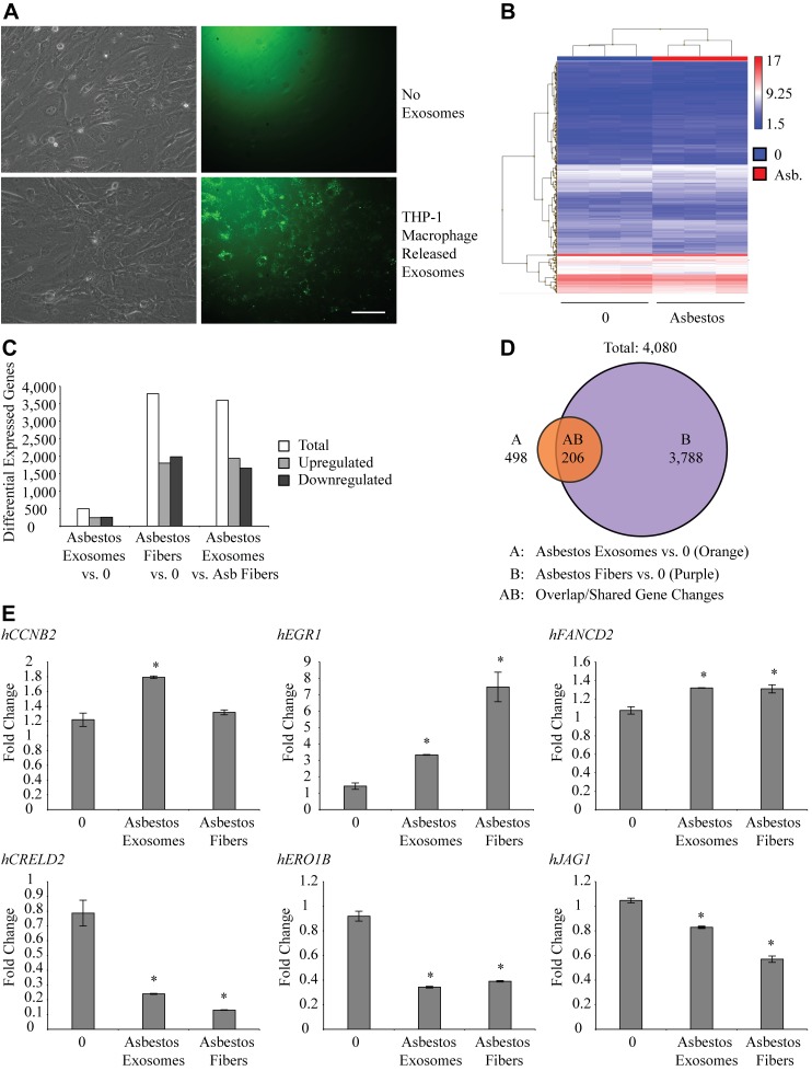
Figure 5.
Exosomes from asbestos-exposed THP-1 cells are taken up by mesothelial cells and caused gene expression changes. A) PKH67 labeled exosomes from THP-1 cells interact with and are taken up by target mesothelial cells. Scale bar, 100 nm. B) Clariom S microarray heat map of gene expression between control mesothelial cells and mesothelial cells exposed to exosomes from asbestos-exposed macrophages. C) Number of differentially expressed genes from microarray analysis in groups of asbestos exosomes vs. control, asbestos fibers vs. control, and asbestos exosomes vs. asbestos fibers. D) Venn diagram showing genes differentially expressed between control mesothelial cells and asbestos exosome exposed cells (A), control- and asbestos fiber–exposed mesothelial cells (B), and the shared genes differentially expressed between both comparisons (A, B). E) qRT-PCR validation of genes up-regulated in asbestos exosome and asbestos fiber groups compared to control (CCNB2, EGR1, and FANCD2) and genes down-regulated in asbestos exosome and asbestos fiber groups compared to control (CRELD2, ERO1B, and JAG1). *P ≤ 0.05, by 1-way ANOVA.

