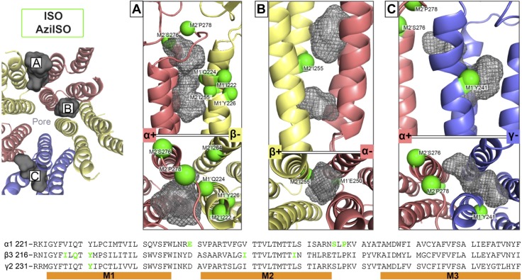
Figure 11.
Docking experiments within predicted ISO binding cavities as indicated by AziISO photolabeled residues in the α1β3γ2L GABAA receptor. The 3 suggested transmembrane domain binding cavities within the α+/β− (A) and β+/α− (B) interfaces and the α+/γ− intrasubunit/intersubunit cavity (C). All residues that face cavities were made flexible, whereas the backbone structure remained rigid during docking experiments. The gray Connolly surface/mesh dotted representations are of 5 ISO and 5 AziISO in the highest scored poses predicted by AutoDockVina (33). Green spheres indicate the Cα atoms of residues photolabeled by AziISO in the α1β3γ2L GABAA receptors and are labeled accordingly. The left panel includes a transmembrane domain view of the docking experiments for each unique interface from the synaptic cleft [only 1 of 2 β+/α− interfaces (B) shown]. The bottom panel includes the aligned α1-, β3-, and γ2L-subunit sequences that span the M1–M3 helices, with photolabeled residues colored in green.

