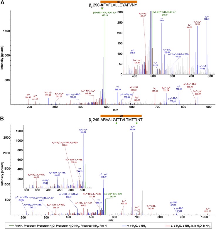
Figure 7.
AziISO photolabeled residues within the β3-subunits of α1β3 and α1β3γ2L GABAA receptors. Mass spectra of β3-subunit AziISO photolabeled residues β3-M3′ V290 (A) and β3-M2′ I255/β3-M2′ I264 (B) within α1β3 and α1β3γ2L GABAA receptors, respectively. Focused view the spectrum within ∼350–825 (A) and 280–590 m/z (B), with b (red), y (blue), and precursor ions (green) shown. Predicted photolabeled residues is shown in bold, underlined, and is indicated by a green asterisk. Above the spectra are the subunit peptide sequences that contain the β3-subunit transmembrane helices. Predicted photolabeled residue is shown in bold, underlined, and is indicated by a green asterisk. See the Supplemental Material for associated peptide fragment tables.

