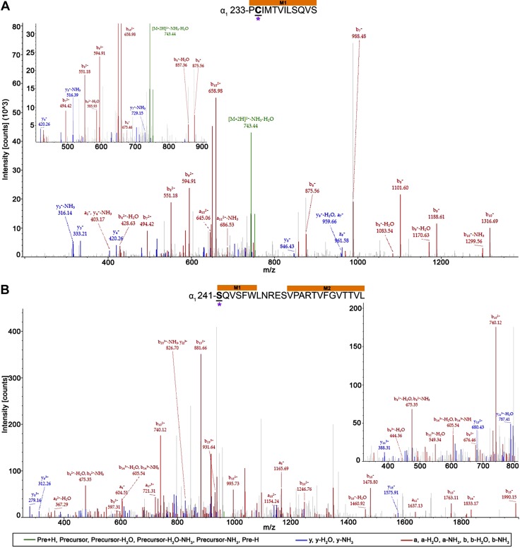
Figure 8.
AziSEVO photolabeled residues within the α1-subunits of α1β3 and α1β3γ2L GABAA receptors. Mass spectra of α1-subunit AziSEVO photolabeled residues α1-M1′ C234 (A) and α1-M1′ S241 (B) within α1β3 and α1β3γ2L GABAA receptors, respectively. Above the spectra are the subunit peptide sequences that contain the α1-subunit transmembrane helices. Focused view of the spectrum within ∼425–900 (A) and 325–800 m/z (B), with b (red), y (blue), and precursor ions (green) shown. Predicted photolabeled residue is shown in bold, underlined, and is indicated by a magenta asterisk. See the Supplemental Material for associated peptide fragment tables.

