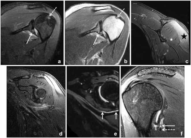
Figure 2.
A 33-year-old woman status post left shoulder dislocation. (a) Oblique coronal inversion recovery and (b) proton density magnetic resonance images of the left shoulder demonstrate a Hill-Sachs lesion (dashed arrow) and partial capsular detachment from the scapula (solid arrow). Dedicated magnetic resonance imaging of the left axillary nerve was acquired at 6-week follow-up to evaluate a dense axillary nerve palsy postdislocation. (c) Coronal T2-weighted Dixon fat-suppressed image confirms denervation edema of deltoid muscle (black star) and relative sparing of the teres minor (white star). (e) Axillary nerve (arrows) is better delineated from adjacent vessels on vascular-suppressed, 3-dimensional T2-weighted curved multiplanar reformatted image compared with (d) the 2-dimensional image without vascular suppression. (f) T2-weighted sagittal image confirms suspected stretch injury, with signal hyperintensity of the axillary nerve (solid arrow) adjacent to the capsule with the posterior circumflex humeral artery (dashed arrow).

