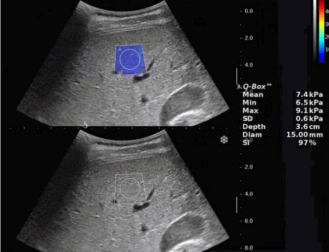
Fig. 1. Elastography of a 14-year-old boy who underwent abdominal ultrasonography for evaluation of glycogen storage disease.

The B-mode image and shear wave elastography (SWE) elastographic images were placed up and down. A SWE box (2.0×2.5 cm sized) was placed in the right anterior segment of the liver, avoiding vascular structures. After the entire area of SWE box was color-coded, 15-mm2 circular region of interest was placed carefully in the area where the color coding was as homogeneous as possible not to include any artifacts related to motion or pulsation.
