
Fig. 6. A 60-year-old woman with quadriceps tendinosis.
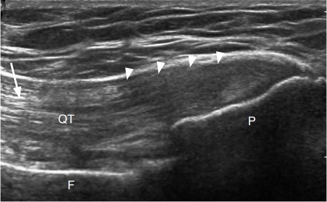
Ultrasonography of the anterior knee long axis to quadriceps tendon (QT) shows a thickened and hypoechoic tendon (arrowheads). Proximally, a normal fibrillar pattern is seen (arrow). P, patella; F, femur.