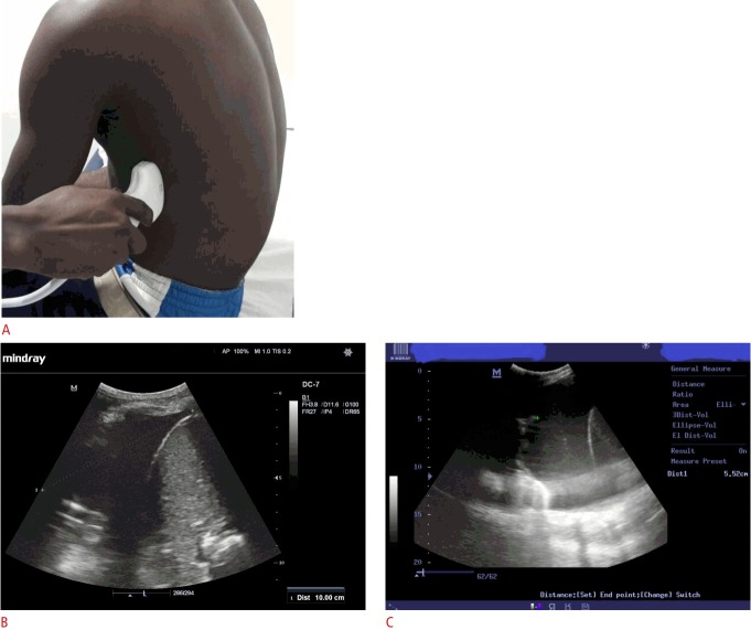
Fig. 2. Right-sided pleural effusion in a 60-year-old man.
A. This image depicts the patient and probe positions for obtaining measurements for the erect formulae. B, C. Corresponding chest sonography shows the craniocaudal extent (cursors) of the effusion (B) at the dorsolateral chest wall (erect 1 formula), as well as the lung base to mid-diaphragm distance/subpulmonary height (C) of the effusion (erect 2 formula).

