Figure 4.

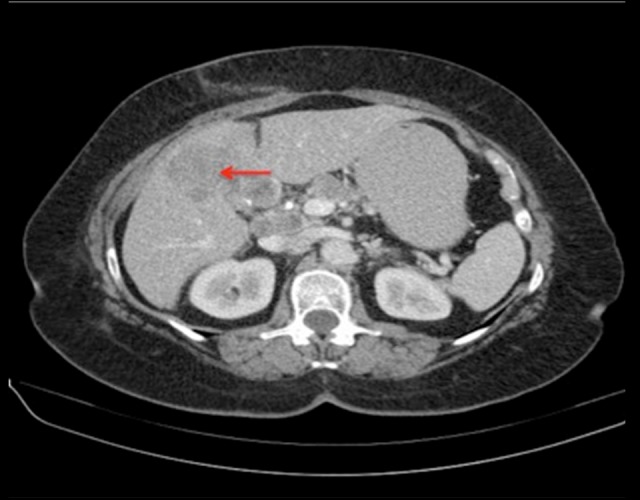
Transaxial CT image demonstrates a large hypoenhancing area in the medial aspect of the left hepatic lobe predominantly involving segment 4B (red arrow). Lymphadenopathy involving the nodes in the porta hepatis is also noted.

Transaxial CT image demonstrates a large hypoenhancing area in the medial aspect of the left hepatic lobe predominantly involving segment 4B (red arrow). Lymphadenopathy involving the nodes in the porta hepatis is also noted.