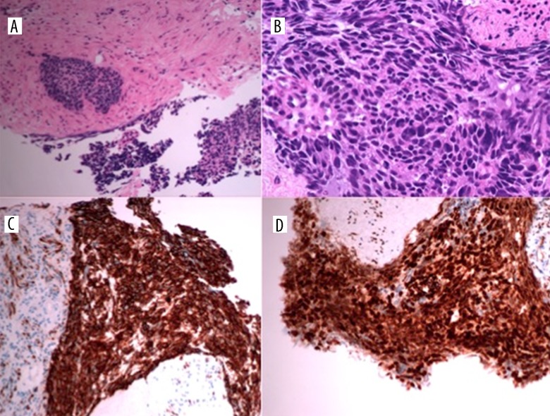
Figure 9.
Histopathological analysis of the pelvis mass biopsy reveals dense sheets of small cells with scanty cytoplasm, hyperchromatic nuclei, frequent mitoses, and inconspicuous nucleoli, consistent with small-cell carcinoma (A, B). On immunohistochemistry, CK AE1/AE3 shows positive staining (C) and WT-1 is also positive (D).

