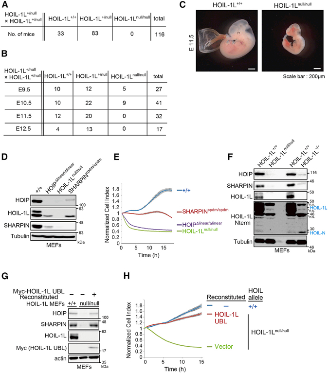
Figure 1. Disruption of HOIL-1L UBL Results in Embryonic Lethality.

(A) Number of offspring of each genotype resulting from crosses of HOIL-1L+/null” mice.
(B) Numbers of embryos obtained at each embryonic stage (E9.5, 10.5, 11.5, and 12.5) from crosses of HOIL-1L+/null” mice.
(C) Representative gross appearance of HOIL-1L-null and WT littermate on embryonic day 11.5 (E11.5).
(D) Immunoblot analyses of lysates of MEFs from mice of the indicated genotypes.
(E) Indicated MEFs were stimulated with TNF-α (10 ng/ml), and cell viability was continuously measured on a real-time cell analyzer (RTCA) (means ± SEM, n = 3).
(F) Immunoblot analysis of lysates of MEFs from mice of the indicated genotypes. ns, non-specific band.
(G) Immunoblot analysis of lysates of HOIL-1L-null MEFs reconstituted with HOIL-1L UBL.
(H) Cell viability of the indicated MEFs was measured as described in Figure 1E (means ± SEM, n = 4).
See also Figure S1.
