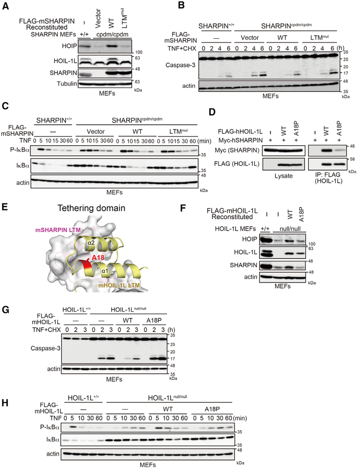
Figure 5. Crucial Role of LTM-Mediated HOIL-1L/SHARPIN Interaction in Trimeric LUBAC Formation and Stabilization.

(A) Immunoblot analysis of lysates from WT or cpdm MEFs reconstituted with the indicated proteins.
(B and C) cpdm MEFs stably reconstituted with the indicated proteins were stimulated with TNF-α (10 ng/ml) plus CHX (20 mg/ml) (B) or TNF-α (10ng/ml)(C) fortheindicated periods, followed by immunoblotting.
(D) The indicated expression plasmids were transfected into HEK293T HOIP KO cells. Cell lysates and anti-FLAG immunoprecipitates were immunoblotted as indicated.
(E) LTMs of HOIL-1L and SHARPIN are shown as a ribbon model and on the molecular surface, respectively. Ala18 of HOIL-1L (red) is located at the surface of the TD.
(F) Cell lysates of HOIL-1L-null MEFs stably expressing the indicated proteins were probed as indicated.
(G and H) HOIL-IL-null MEFs stably expressing the indicated proteins were stimulated with TNF-α (10 ng/ml) plus CHX (20 mg/ml) (G) or TNF-α (10 ng/ml) (H) for the indicated periods, and cell lysates were analyzed by immunoblotting.
See also Figure S5.
