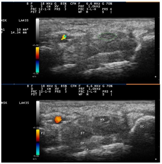
Figure 1.
Ultrasound image of the median nerve at the carpal tunnel inlet. The image shows the median nerve outlined using a free trace method inside the hyperechoic rim of the nerve.
Abbreviations: CP, carpal bones (lunate); DP, deep tendons (flexor digitorum profundus); MN, median nerve; PI, pisiform bone; SC, scaphoid bone; ST, superficial tendons (flexor digitorum superficialis); UA, ulnar artery.

