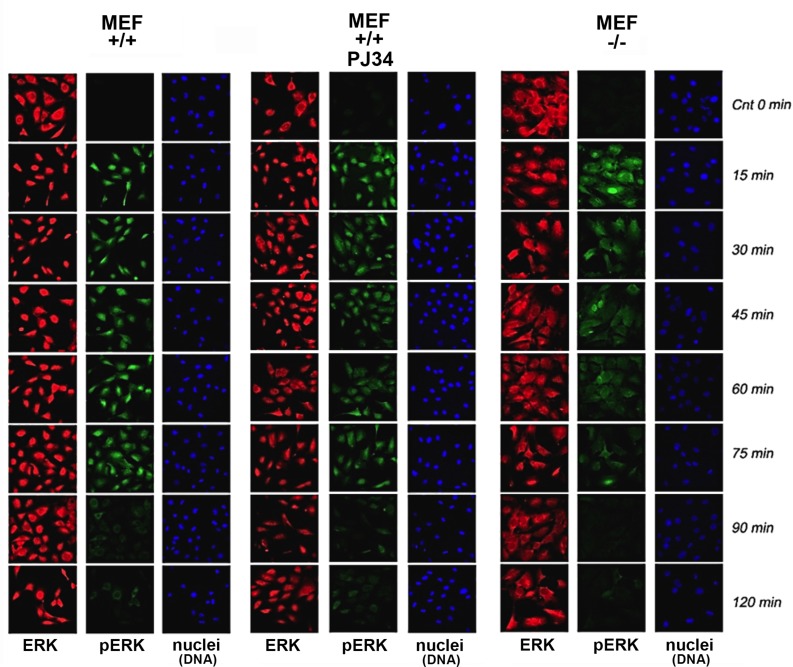
Figure 1. Stimulation–induced Erk phosphorylation down-regulated in nuclei of PARP1-KO MEF.
Immunolabeled Erk (red) and phosphorylated Erk (green) monitored by confocal microscopy during 120 min after stimulation with PMA (200 ng, 15 min) in nuclei of MEF, without and after PARP1 inhibition, and in nuclei of PARP-KO MEF. DNA is labeled with DAPI (blue).

