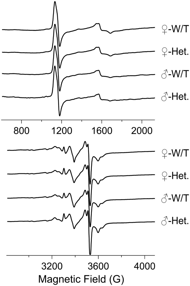
FIGURE 4.
Electron paramagnetic resonance spectra from muscle tissue. The traces show EPR spectra from quadriceps muscle samples from female wild-type (n = 5), female Sdha+/- (n = 7), male wild-type (n = 4), and male Sdha+/- (n = 2) rats. The EPR signals in the 4 groups do not display any statistically significant differences. Data were analyzed using the Student t-test.

