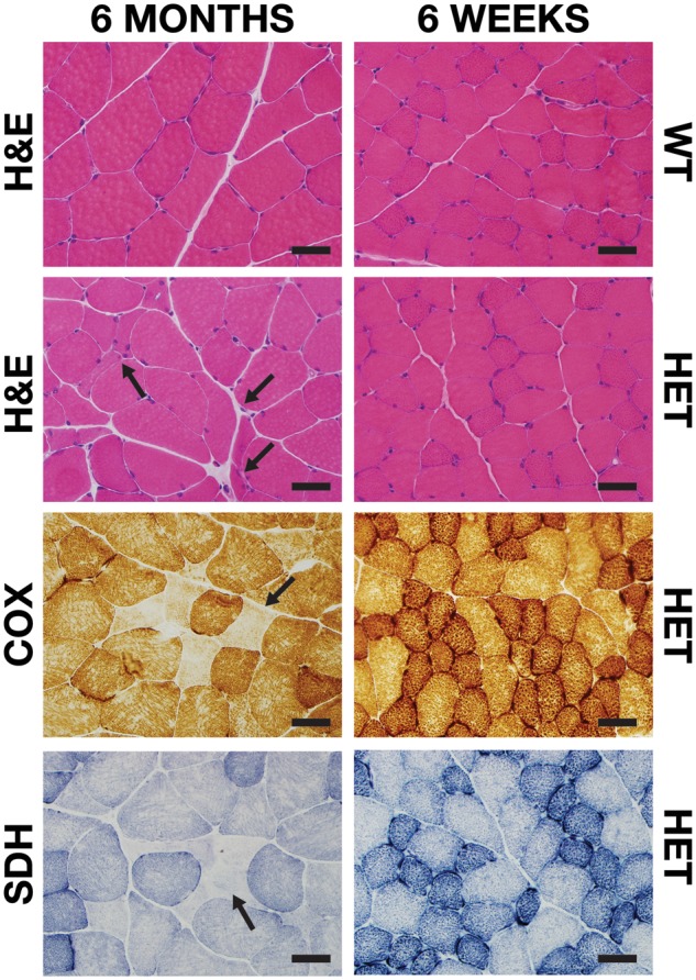
FIGURE 5.

Histological staining of Sdha+/- muscle. Representative areas of transversely sectioned gastrocnemius muscle after sectioning and staining. Upon H&E staining, mild fiber atrophy (arrows), basophilic fibers, and internal nucleation were observed in 6-month-old Sdha+/- (HET) animals. COX and SDH staining revealed rare, atrophic, COX and SDH-deficient fibers (arrows) in 6-month-old Sdha+/- animals. Scale bar = 40 µm.
