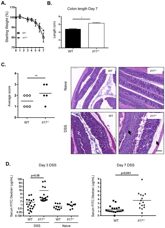
Figure 1. Il-17−/− mice suffer worse epithelial injury and enhanced gut permeability after DSS administration.
(A) Weight loss of WT (squares), Il-17−/− (triangles) mice over time during DSS treatment, representative data from 3 independent experiments, n=5–7/group, mean ± SEM. (B) Colon length at day 7, combined data from 3 experiments, mean ± SEM. *p<0.05, **p<0.001 (C) Il-17−/− colons exhibit increased bleeding into lumen, representative colons from 3 experiments, n=4–7/group. (D) Pathology scores of disease severity. H&E of colons reveal enhanced edema and lymphocytic infiltrate in Il-17−/− colons (arrows) after DSS, 20x magnification shown. (E) Detection of FITC-dextran in plasma showing increased colon permeability in Il-17−/− over WT after 3 and 7 days of DSS, representative data from 2–3 experiments, or combined data from 3 experiments (day 7), means indicated, *p<0.01. All statistics generated using the one-way ANOVA, with Tukey’s Multiple Comaprisons.

