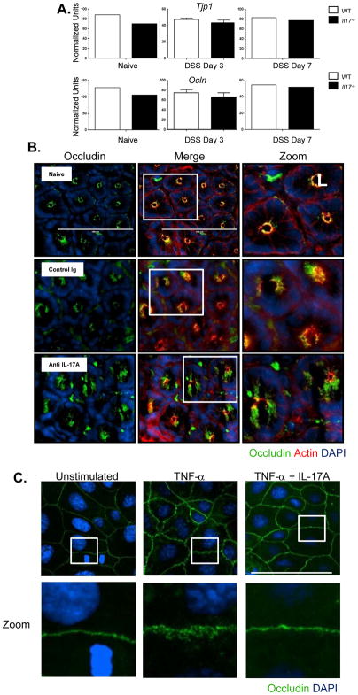
Figure 2. Dysregulation of occludin cellular localization in the absence of IL-17A following DSS induced injury.
(A) Colonic tissues were analyzed by RT PCR for the tight junction proteins ZO-1 and occludin mRNA at day 3 and day 7 after DSS treatment. No difference in ZO-1 or occludin message between WT or Il-17 −/− mice. (B) C57BL/6 mice were subcutaneously administered with either control IgG or anti-IL-17A antibody at 20mg/kg 2 days before DSS treatment. Immunofluorescence images of occludin (green), f-actin (red), DNA (blue) of distal colon segments 3 days after DSS. The fluorescent images depict cross sections of the intestinal crypts in the distal colon with the apical surface of the cell oriented toward the lumen (L). The third column represents a magnified image from the white box in the second column. (C) Caco-2 cells were plated on Poly-L-lysine coated coverslips and treated with recombinant human TNFα (10ng/mL) or TNFα (10ng/mL) + recombinant human IL-17A (10ng/mL) and cultured for 24 hrs. Immunofluorescence images of occludin (green) and DNA (blue). The bottom row represents a magnified image from the corresponding white box in the first row.

