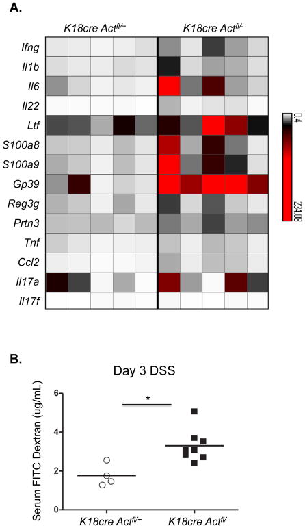
Figure 3. Enhanced inflammatory signature in the absence of IL-17-induced Act-1 signaling in epithelial cells.
(A) Epithelial cells from the distal colons of control or K18CreAct1fl/− DSS-treated mice were analyzed by RT-PCR. Message levels of inflammatory genes are represented in a heat map as fold change over levels in naïve mice. Each column represents an individual mouse; n=5/group. (B) Detection of FITC-dextran in plasma after 3 days of DSS-treatment revealing increased permeability in epithelial specific Act-1 deficient mice. One of two experiments is shown; *p value = 0.01.

