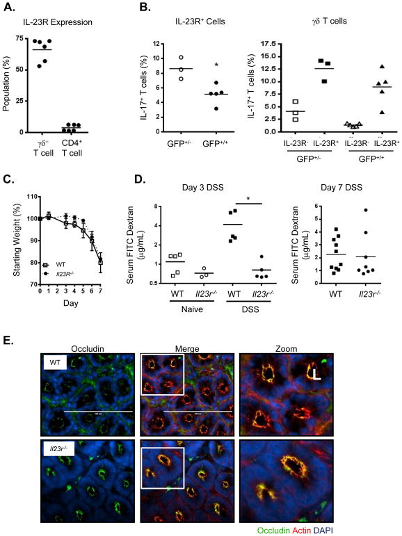
Figure 6. IL23R deficient mice do not exhibit increased gut permeability after DSS treatment.
(A) IL-23R expression in different lamina propria lymphocyte populations as determined by Il-23r-Gfp reporter using Il-23r-Gfp+/− mice, combined data from two experiements. (B) IL-17 production is decreased overall in the absence of IL-23R signaling as shown by comparison of Il-23r-Gfp+/− (GFP+/−) (IL-23R present) and Il-23r-Gfp+/+ (GFP+/+ )(IL-23R absent) mice for all GFP+ cLPL (left panel) or γδ T cells only (right panel). (C) Weight loss of WT (squares), Il-23r −/− (circles) mice over time during DSS treatment, representative data from 3 independent experiments, n=5–7/group, mean ± SEM. (D) Detection of FITC-dextran in serum showing increased colon permeability in WT over Il23r −/− mice after 3 and 7 days of DSS, representative data from 2–3 experiments, or combined data from 2 experiments (day 7), means indicated, *p<0.01. All statistics generated using the one-way ANOVA, with Tukey’s Multiple Comaprisons. (E) Immunofluorescence images of occludin (green), f-actin (red), DNA (blue) of distal colon segments from WT or Il23r −/− mice 3 days after DSS. The fluorescent images depict cross sections of the intestinal crypts in the distal colon with the apical surface of the cell oriented toward the lumen (L). The third column represents a magnified image from the white box in the second column.

