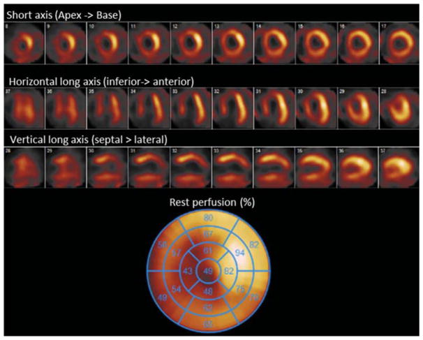
Figure 2.
SPECT rest only myocardial perfusion imaging with 99mTc-sestamibi for viability assessment of a patient with severe three vessel disease and LV EF 35%. Visual and quantitative analysis reveals a large region of infarct in the apical to mid anterior, anteroseptal and inferoseptal segments. The quantitative polar plot shown reveals viability in all coronary territories except in segments of the apex with perfusion under 50%.

