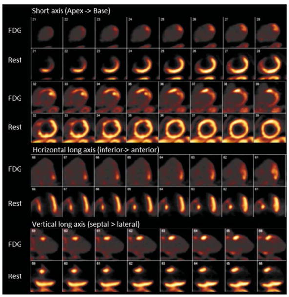
Figure 5.
PET myocardial perfusion imaging using N-13 ammonia rest perfusion images (bottom row images) and F-18 fluorodeoxyglucose (FDG) myocardial metabolic images (top row images). A large fixed perfusion defect with akinesis on gated images is seen in the mid to basal anterior and septal segments of the LAD territory. No FDG uptake is seen in this territory suggesting no viability.

