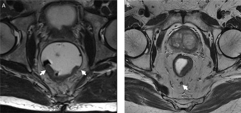
Figure 1.

Neoadjuvant-eligible staging findings using pelvic magnetic resonance imaging. A) T3 midrectal lesion with posterior invasion through the muscularis propria (low intensity signal encircling rectum) into the mesorectal fat with arrows marking lateral extent. B) T2 mid-rectal, posterior lesion with thinning of the muscularis propria and a 5mm pathologic lymph node (arrow) located in the posterior mesorectal fat.
