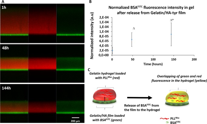
Figure 5.
Diffusion of BSAFITC from the film (2D) to the hydrogel (3D). (A) Confocal images of the two-layer system at different time points, with the gelatin/HA-tyr cross-linked film layer labeled with BSAFITC with a 6% w/v gelatin hydrogel cross-linked with a 20% w/v TGA solution labeled with PLLRho (red fluorescent probe) on top of it. Starting from 1 h, the diffusion of BSAFIT from the film to the hydrogel was monitored (from left to right, the green and red channels are merged, the red channel to localize the hydrogel labeled with the red fluorescent probe and the green channel to localize the film with BSAFITC initially loaded inside). Overtime, we can see the green fluorescence (BSAFITC) initially localized in the film (bottom part) moving to the hydrogel (upper part in red), which means that BSA has diffused from the film to the hydrogel. (B) Quantification of the green fluorescence (BSAFITC) in the hydrogel at different times normalized by the area of the hydrogel (p ≤ 0.05). (C) Schematic representation of the experimental setup.

