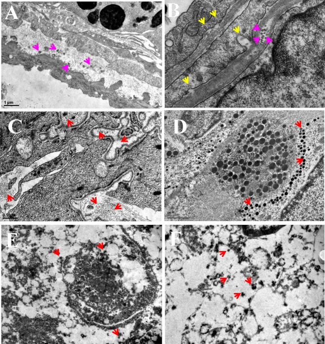
Figure 7.
In vitro cellular uptake of PMS nanocomposites by MDA-MB-231 cells witnessed the accumulation of nanocomposites in the cytoplasm and nucleus. (A) and (B) TEM image reveals PMS nanocomposites get internalized into the cell membrane through receptor-mediated endocytosis. (C) and (D) TEM images of PMS nanocomposites distributed within the cancer cells. (E) and (F) The PMS nanocomposites distributed throughout the nucleus. Red arrows point out the distribution of spherical shaped PMS nanomaterials. Scale bar = 1 μm.

