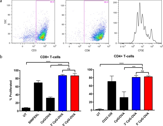
Figure 4.
Proliferation of CD8 and CD4 T-cells following 5′ and 3′ CpG–OVA conjugate treatment. BMDCs were pulsed with a mixture of CpG (0.2 μM) and OVA (79 nM), 3′ CpG–OVA conjugate (equivalent to 0.2 μM CpG and 79 nM OVA), 5′ CpG–OVA conjugate (equivalent to 0.2 μM CpG and 79 nM OVA), or a control of either SIINFEKL (2.6 μM) or OVA323-339 (1.4 μM) peptide. Sorted, CFSE-stained CD8+ or CD4+ T-cells were co-cultured with pulsed BMDCs at a ratio of 1:10. (a) Gating strategy to identify the proliferation peaks of CD8 T-cells. (b) Percent proliferated CD8+ and CD4+ T-cells after incubation with activated BMDC for 72 h. Bars represent the mean of three independent experiments ± SEM; statistical significance was determined by one-way ANOVA Dunnett’s post hoc test; ****p < 0.0001, ***p < 0.001, ns = nonsignificant.

