Figure 1.

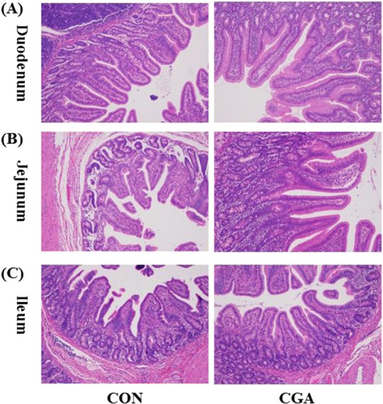
Histological evaluation of duodenal (A), jejunal (B), and ileal (C) tissues (H & E; ×100) after exposure to CGA. CON, pigs receiving a basal diet; CGA, pigs receiving a basal diet supplemented with 1000 mg/kg CGA.

Histological evaluation of duodenal (A), jejunal (B), and ileal (C) tissues (H & E; ×100) after exposure to CGA. CON, pigs receiving a basal diet; CGA, pigs receiving a basal diet supplemented with 1000 mg/kg CGA.