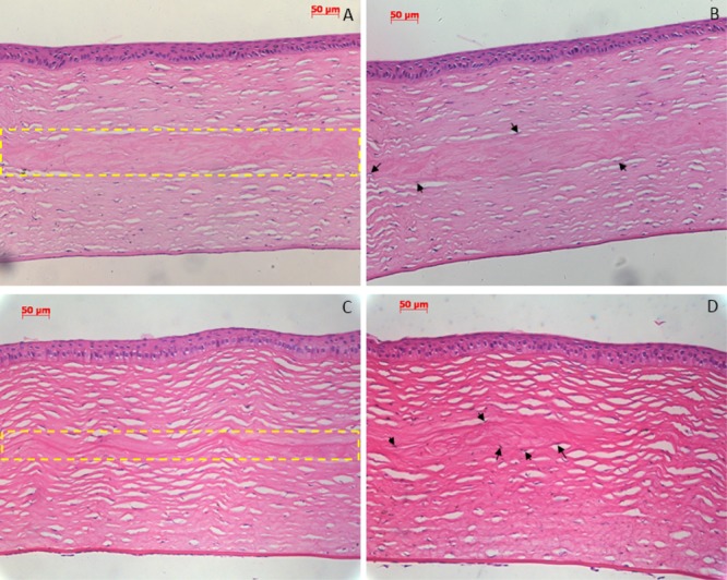
Figure 5.

H&E staining sections of the rabbit cornea after implantation. (A,B) Postoperative 5 months: there was good biocompatibility and connection between the implant and stroma [(A) inside the yellow dashed frame]; no inflammatory cells and new vessels were observed; and some keratocytes began to grow into the superficial lamellae of implant (B). (C,D) Postoperative 7 months: implant was almost degraded without causing adverse inflammatory or immune reactions [(C) inside the yellow dashed frame], only a little residuary implant was observed, and some keratocytes began to grow into the superficial lamellae of the implant (D). Arrow indicates new keratocytes grown into the superficial lamellae of the implant.
