Figure 1.
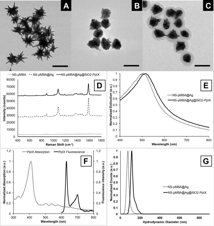
TEM images of gold nanostars (A), pMBA-embedded silver-coated gold nanostars (B), and silica-coated, pMBA-embedded silver-coated gold nanostars (C; scale bars are 100 nm). (D) SERS signal from pMBA-labeled gold nanostars (dotted), the silver-embedded pMBA-labeled gold nanostars (dashed), and the silica-coated silver-embedded pMBA-labeled gold nanostars (solid); 10 s acquisition, 0.65 mW laser power. (E) UV/vis extinction spectra of the silver-coated nanostars before (dotted) and after (solid) silica coating. (F) PpIX absorption (dotted) and emission (solid) at 415 nm excitation. (G) Particle size distribution of the silver-coated gold nanostars before (dotted) and after (solid) silica coating.

