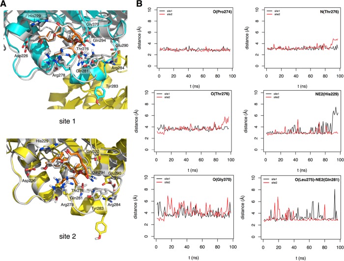
Figure 4.
Model of dictyostatin bound to a MT. (A) Superimposition of a representative structure of each major cluster of dictyostatin conformers (C atoms colored in orange) bound to β-tubulin in subunit B (C atoms colored in blue, site 1) and F (C atoms colored in yellow, site 2) along the MD simulation (100 ns) onto subunit B of the crystal structure of the dimeric tubulin in the complex with dictyostatin (C atoms colored in gray) reported in this work. (B) Time evolution of distances (Å) relevant to the pharmacophore along the MD simulation in the sites of B and F tubulin monomers.

