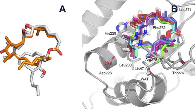
Figure 5.

Comparison between the predicted structures of the analogues. (A) Superimposition of a representative structure of the major conformer of isodictyostatin (2, C atoms colored in gray), as simulated using MD for 100 ns in a box of TIP3P water molecules, onto the crystal structure conformation of dictyostatin (dictyostatin, C atoms colored in orange). (B) Molecular model showing the best docking poses of compounds 4–16 bound to the taxane-binding site of β-tubulin. The very weak binder isodictyostatin (2) and hybrid 14, which incorporates a fragment derived from docetaxel, are not shown.
