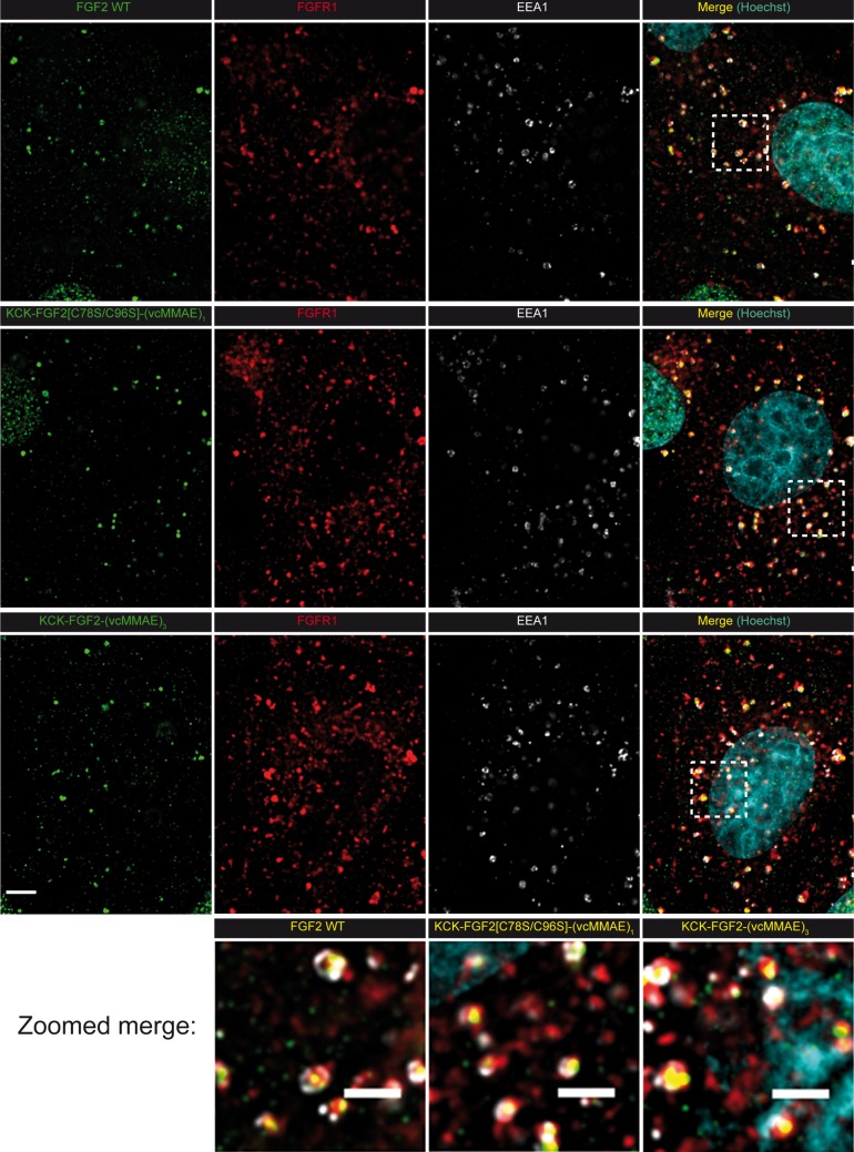
Figure 6.
Widefield immunofluorescence microscopy imaging of endocytosed FGF2 WT and FGF2-vcMMAE conjugates in U2OS-R1 cells. U2OS-R1 cells cultured on glass coverslips were incubated with 500 ng/mL FGF2 WT, KCK-FGF2[C78S/C96S]-(vcMMAE)1, or KCK-FGF2-(vcMMAE)3 at 37 °C for 40 min to allow for endocytosis. The cells were stained with anti-FGF2 (green), anti-FGFR1 (red), and anti-EEA1 (white) antibodies and with Hoechst 33342 to visualize DNA. Images were deconvolved and single optical sections are shown, either as single-channel (color) images or as overlays as indicated. The bar corresponds to 4 μm, and in zoomed images, to 2 μm. The squares marked in four-color overlay images indicate blown-up regions shown in the bottom row.

