Figure 3.

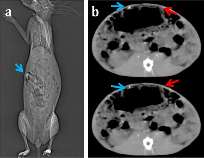
(a) In vivo plain radiography of CDI sutures in rabbit and (b) CT images of CDI implants (blue arrows) and plain gut sutures (red arrows) implanted at a symmetrically opposite location.

(a) In vivo plain radiography of CDI sutures in rabbit and (b) CT images of CDI implants (blue arrows) and plain gut sutures (red arrows) implanted at a symmetrically opposite location.