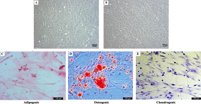
Figure 5.
BMSC morphology (100×), cell adherent growth, spindle and polygonal, (A) P0, (B) P3. (C) Intracellular oil red O staining indicating lipid-rich vacuole formation of the rb-BMSCs after three weeks of adipogenic induction. (D) Alizarin red staining demonstrated that the mineralized nodules formed in the BMSCs after three weeks under the osteogenic induction. (E) After four weeks of chondrogenic induction, the cell was sectioned and stained with toluidine blue; the positive acidic proteoglycan indicated the chondrocyte-like cell formation. Scale bar = 100 μm.

