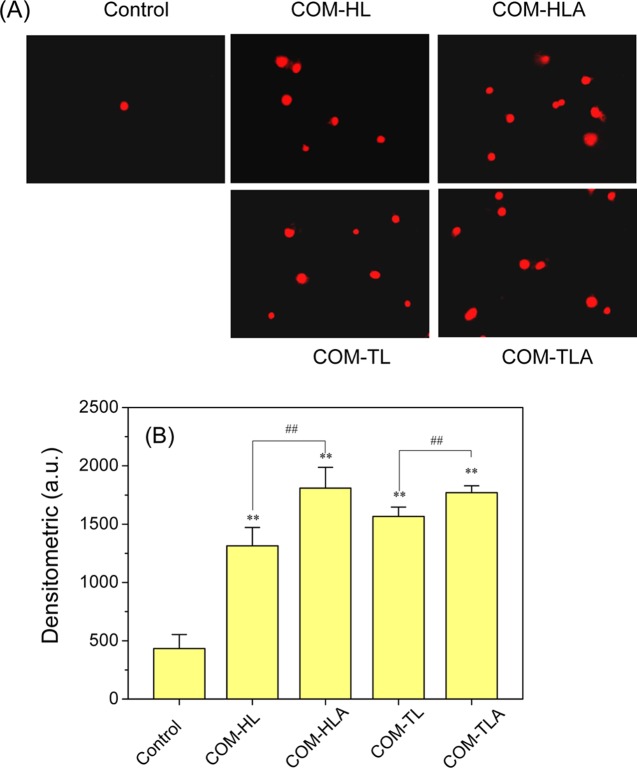
Figure 5.
Fluorescence images of HK-2 cells stained by PI (A) and quantitative analysis results of red fluorescence intensity (B) after exposure to 400 μg/mL COM crystals in various shapes for 6 h. Scale bar: 50 μm. Compared with control group, *p < 0.05, **p < 0.01. COM-HL treatment group vs COM-HLA treatment group, COM-TL treatment group vs COM-TLA treatment group, #p < 0.05, ##p < 0.01.

