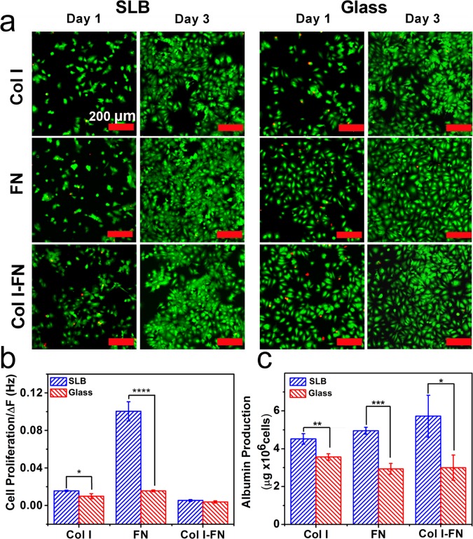
Figure 5.
Cell viability and metabolic functions on ECM-coated platforms. (a) Representative fluorescence micrograph images of detecting live (green) and dead (red) Huh 7.5 cells cultured on SLB and glass coated with Col I and FN and Col I–FN at 1 and 3 days post seeding. Scale bar is 200 μm. (b) Proliferation of Huh 7.5 cells was measured over a 3 day period. The values were normalized to the absolute frequency shifts due to the amount of adsorbed proteins. (c) Albumin production by Huh 7.5 cells plated on different platforms measured after 24 h of cell growth. The values were normalized to the total number of cells (n = 3, mean ± SD, *P ≤ 0.05, **P ≤ 0.01, ***P ≤ 0.001, and ****P ≤ 0.0001).

