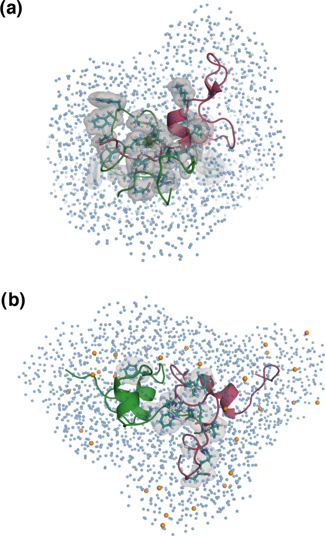
Figure 6.

Representative structures from the dimeric ensembles (a) PW-D and (b) PG-D; the two peptide units are colored in pink and green. The residues involved in interpeptide high probability contacts are depicted by a translucent gray surface, with side chains represented as sticks and colored teal. Glucose molecules around the PG-D dimer within a distance of 7 Å from the protein units are shown as orange colored spheres and the water oxygens around the dimers in both the systems are shown as spheres colored skyblue.
