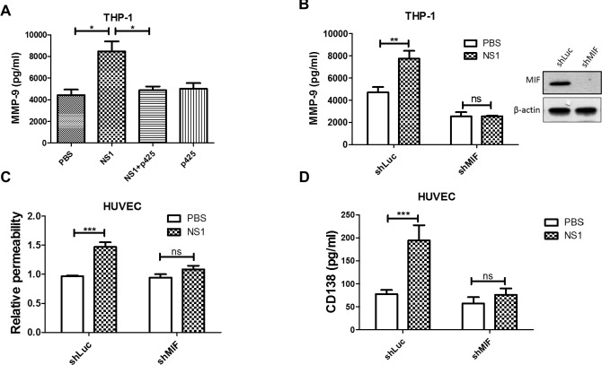
Fig 6. NS1-induced MMP-9 secretion from THP-1 cells is regulated by MIF.
(A) PMA-activated THP-1 cells were treated with PBS, NS1, or NS1 with p425 for 24 h, and the concentration of MMP-9 in the supernatant was determined by ELISA (n = 5). (B) The MMP-9 levels in the supernatants of PMA-activated THP-1-shLuc and THP-1-shMIF cell cultures were detected by ELISA after incubation with or without NS1 for 24 h. (n = 3) (C) PMA-activated THP-1-shLuc and THP-1-shMIF cells were incubated with PBS or NS1 for 24 h; then, the supernatant was collected and incubated with HUVEC monolayers grown in upper Transwell chambers. After 24 h of incubation, endothelial permeability was determined using streptavidin-HRP and TMB. (n = 6) (D) The supernatant of THP-1-stimulated HUVEC cultures was collected, and the CD138 concentration was determined by ELISA (n = 5). *P<0.05, **P<0.01, ***P<0.001; ns, not significant; Kruskal-Wallis ANOVA (panel A), unpaired t-test (panel B, C and D).

