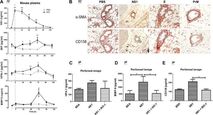
Fig 7. MIF inhibition attenuates DENV NS1-induced glycocalyx degradation in mice.
(A) Before the injection of PBS or NS1, the blood of 8- to 12-week-old BALB/c mice (n = 3) was collected by orbital sinus sampling with 10% citrate. Next, the mice were intravenously injected with 50 μg of NS1 or 100 μl of PBS. Blood samples were collected from the mice immediately after injection and every 24 h thereafter until 120 h. The plasma concentrations of NS1, MIF, HPA-1, and MMP-9 were measured by ELISA. Arrow, injection time point. (B) BALB/c mice received two subcutaneous injections of PBS, NS1, or recombinant E or prM at the same location within 24 h. One day after the second injection, the mice were sacrificed, and a series of skin tissue sections were hybridized with anti-α-SMA and anti-CD138 antibodies and stained with DAB (brown). (C-E) BALB/c mice (n = 5) were intraperitoneally injected with BSA or NS1 with or without an MIF inhibitor (ISO-1), and the peritoneal lavage fluid was collected 24 h after the injection. The concentrations of (C) HPA-1, (D) MMP-9 and (E) CD138 in the peritoneal lavage fluid were measured by ELISA. *P<0.05, **P<0.01, ***P<0.001; unpaired t-test (panel A), Kruskal-Wallis ANOVA (panel C, D and E).

