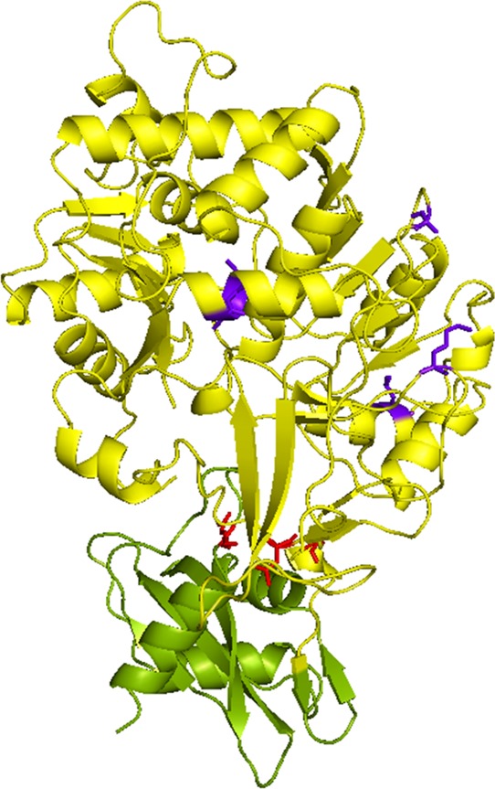
Figure 4.

Locations of the introduced mutations depicted in the reported crystal structure of the Ppy luciferase. Protein Data Bank file 1BA3 was obtained and analyzed by PyMOL. The N-terminal domain is shown in yellow, and the C-terminal domain is shown in green. The amino acid mutations derived from LGR and Mutant E are indicated in red and purple, respectively.
