Figure 5.
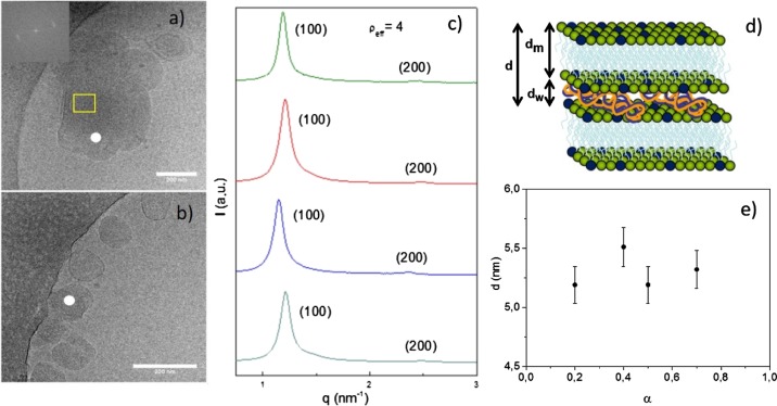
(a,b) Cryo-TEM micrographs of C6C22C6/DOPE–pDNA lipoplexes at ρeff = 4 and molar compositions of the cationic lipid in the mixed lipids of (a) α = 0.2 and (b) α = 0.5. The inset in (a) shows the diffraction spot from the FFT calculations over the selected area on the original micrograph. The white points indicate the lamellar structure with a multilamellar pattern. The scale bar is 200 nm. (c) SAXS diffractograms of C6C22C6/DOPE–pDNA lipoplexes at an effective charge ratio of ρeff = 4 and different molar compositions (α). (d) Three-dimensional scheme of the Lα multilamellar lyotropic liquid-crystal structure. (e) Plot of interlamellar distance (d) of this Lα multilamellar structure as a function of molar composition of the mixed lipids (α) at ρeff = 4.

