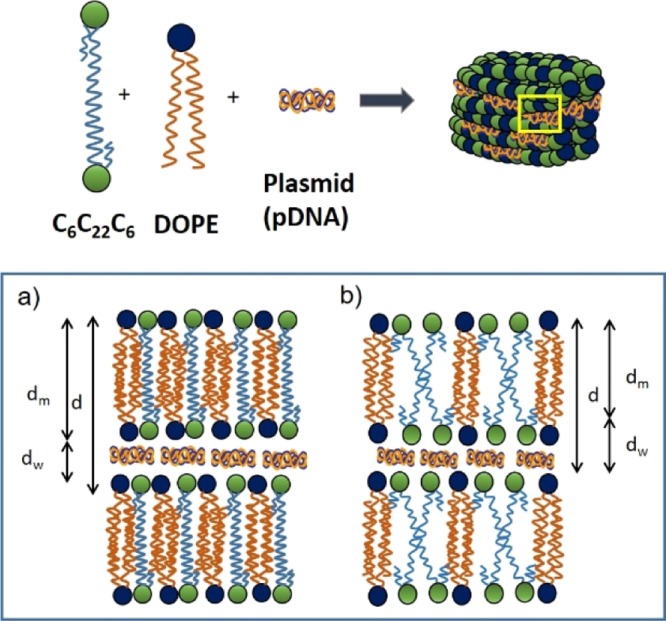
Figure 6.

Schematic drawings of the cationic GBA lipid (C6C22C6), zwitterionic helper lipid (DOPE), and plasmid pDNAs (pEGFP-C3 or pCMV-Luc) self-organized in a Lα multilamellar lyotropic liquid-crystal structure. The inset at the bottom of the figure shows a magnified view of the yellow squared zone of the lipoplex, with two possible arrangements compatible with the SAXS and cryo-TEM results: (a) the 22C spacer of C6C22C6 forms a lipid monolayer and (b) C6C22C6 is organized in a typical lipid bilayer, with the spacer oriented inward and the bilayer with a V shape. In both cases, DOPE is organized in a lipid bilayer fashion, with the hydrophobic chains displaying extensive overlapping to fit the dimensions determined for the interlamellar distance (d).
