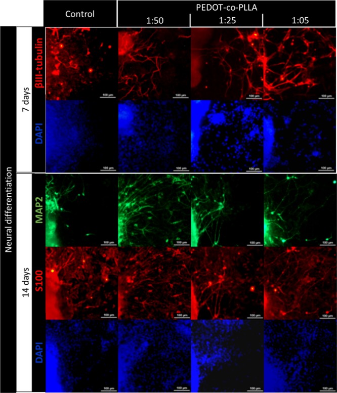
Figure 7.

Characterization of E14.tg2a ESC neural differentiation on a commercial plastic plate (control) and on PEDOT:PDLLA conductive polymers at proportions of 1:50, 1:25, and 1:05. Immunostaining for young neurons with anti-βIII tubulin (red) after 7 days from the beginning of the neural differentiation process. Immunostaining for mature neurons with anti-MAP2 (green) and glial cells with anti-S100β (red) after 14 days. Cell nuclei were stained with DAPI (blue). Scale bar: 100 μm.
