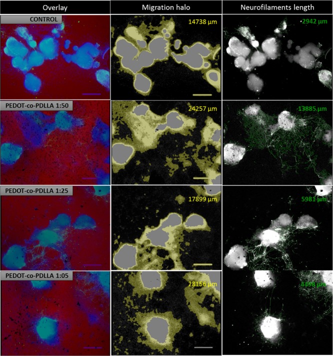
Figure 8.

Quantification of neuronal-differentiated ESCs following anti-MAP-2 staining by StrataQuest analysis software (TissueGnostics). Images of phase-contrast and DAPI and Alexa488 fluorescent channels were overlaid (column 1); migration halo analysis by combining and processing images of DAPI and phase-contrast ROI (column 2); measurement of the total lengths of anti-MAP2-tagged neurites (column 3).
